
An international biophysics team led by Johannes Gutenberg University Mainz and the Max Planck Institute of Biophysics has resolved how nuclear pore complexes protect genomic DNA, proving on November 11, 2026, that the central channel barrier acts as a dynamic fluid rather than a rigid hydrogel. The discovery combines live-cell optical microscopy with exascale supercomputing to resolve a decades-long debate in cellular biology.
Resolution of the Nuclear Pore Permeability Debate
In a study published in Nature on November 11, 2026, researchers demonstrated that intrinsically disordered proteins inside the central channel of human nuclear pore complexes maintain a high-mobility, liquid-like state within intact living cells. The project was co-led by Professor Edward Lemke of Johannes Gutenberg University Mainz and the Institute of Molecular Biology Mainz, alongside Professor Gerhard Hummer of the Max Planck Institute of Biophysics in Frankfurt am Main, with co-first authors Professor Miao Yu from Wuhan University and Dr. Maziar Heidari from the Max Planck Institute of Biophysics.
Nuclear pore complexes serve as the sole gateway perforating the double-membrane nuclear envelope protecting the eukaryotic genome. Each human cell hosts thousands of these structures, regulating the transit of tens of thousands of molecules per minute. Small molecules diffuse passively, but larger proteins and ribonucleic acids require nuclear transport receptors. For over two decades, biophysicists lacked methods to observe barrier proteins in living cells, producing conflicting models in textbook cell biology.
As documented in the Johannes Gutenberg University Mainz press announcement, the team coupled bioorthogonal fluorescent probes directly to phenylalanine-glycine-rich nucleoporins (FG-NUPs) lining the nanocanal. Using time-resolved fluorescence anisotropy and fluorescence lifetime imaging microscopy, the researchers tracked protein movements across a two-hour window. Rather than forming a static obstruction, the protein chains exhibited continuous, nanosecond-scale mobility.
Biophysical Mechanisms: Glycosylation, Importins, and Edge Transport
The investigation explains why isolated nucleoporins behave differently outside cells. When purified in vitro, FG-NUPs aggregate within two hours into a rigid gel resembling gummy confectionery, mirroring pathological aggregates seen in neurodegenerative disease. In living cells, the researchers identified two biochemical factors that prevent gelation and maintain fluidity.
First, the barrier proteins carry extensive post-translational O-linked beta-N-acetylglucosamine (O-GlcNAcylation) sugar modifications. These sugars introduce steric shielding that prevents hydrophobic cross-linking between neighboring motifs. Second, soluble nuclear transport receptors, particularly importin-beta, continuously traverse the channel. By forming rapid, low-affinity multivalent contacts with FG repeats, importin-beta molecules act as dynamic chaperones, preserving liquid dynamics.
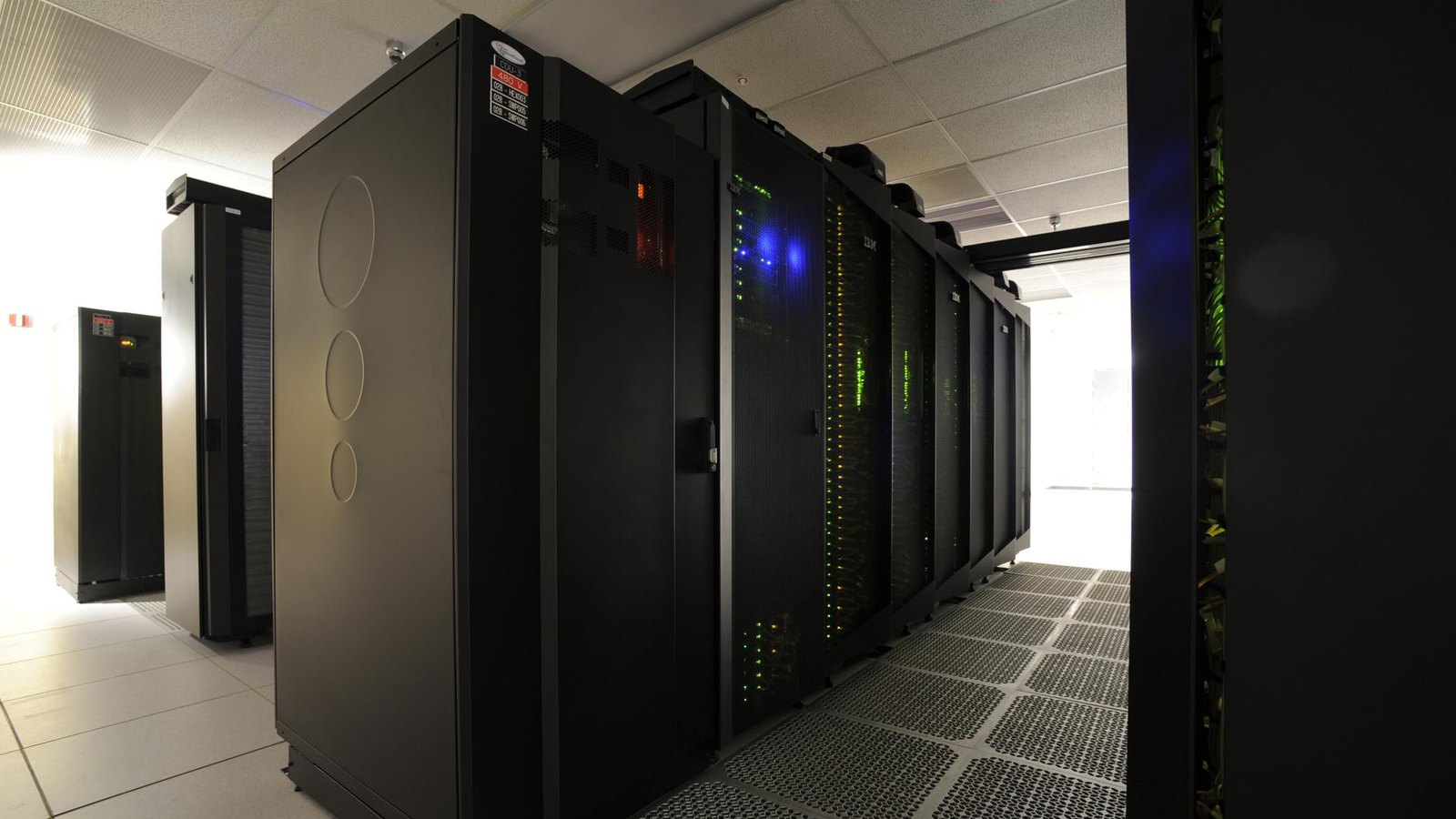
To evaluate these dynamics at molecular scale, the Max Planck team carried out multi-scale molecular dynamics simulation runs on JUPITER, Europe’s first exascale supercomputer at Forschungszentrum Jülich. Modeling a complete nuclear pore complex with over 40 million particles, the calculations confirmed that active transport runs predominantly along the outer periphery of the pore nanocanal rather than through the center, driven by nucleoporin anchor geometry and receptor availability.
| Biophysical Parameter | Historic In Vitro Model | Live-Cell Fluid Discovery (2026) |
|---|---|---|
| Physical State of FG Barrier | Static, cross-linked rigid hydrogel | Highly mobile, nanosecond-scale fluid |
| Role of Sugar Modifications | Omitted from structural models | Essential O-GlcNAcylation prevents self-aggregation |
| Transport Receptor Impact | Local mesh disruption during passage | Constitutive importin-beta flux liquefies barrier |
| Primary Translocation Route | Channel center via transient dilation | Pore periphery and cylinder edge along anchor points |
| Temporal Stability | Solidifies into gummy mass within 2 hours | Maintains stable liquid mobility across cell lifespan |
Context and Historical Controversy Over Nuclear Transport
How nuclear pore complexes combine strict size exclusion with rapid translocation has sparked dispute since the early 2000s. One school argued for a selective phase hydrogel model, wherein hydrophobic bonds create an interlinked mesh that only transport receptors dissolve. A competing school advocated an entropic polymer brush model, claiming thermal flailing of unstructured chains repels macromolecules.
According to an analysis by SciTechDaily report, empirical consensus stalled because X-ray crystallography and cryo-electron microscopy resolve static scaffold rings while blurring flexible disordered domains. By measuring fluorescence lifetimes in intact cells, the Mainz and Frankfurt researchers captured native conformational dynamics.
The study reconciles earlier models: the barrier forms an interconnected network, but cellular glycosylation and receptor flux prevent it from solidifying. Resolving this controversy highlights the necessity of computational reproducibility when pairing exascale supercomputing ensembles with live-cell optical measurements.
Research Community Reaction and Implications for Cellular Aging
Biophysicists have welcomed the study for demonstrating how cells maintain functional phase separation without triggering pathological aggregation. An announcement from the Max Planck Society release highlighted that understanding nucleoporin liquidity informs basic research into cellular defenses against protein misfolding.
“For the first time, we have been able to measure intrinsic protein dynamics within the nanocanal of a living cell and demonstrate that the interior of this canal is fluid-like,” stated Professor Edward Lemke. Lemke noted that when isolated in test tubes without physiological modifiers, nucleoporins turned into a rigid mass, confirming that living cells actively suppress protein hardening.
Professor Gerhard Hummer underscored the role of exascale computing in identifying the peripheral route. “These complex calculations were made possible by JUPITER, Europe’s first exascale supercomputer at the Jülich Research Center,” Hummer explained, noting that simulations show edge-directed transport requires no central physical plug. Analysis from an AlphaGalileo research dispatch noted that nucleocytoplasmic transport failure and nucleoporin aggregation are recognized features of amyotrophic lateral sclerosis (ALS) and Huntington’s disease.
What It Means for Research Facilities and Computational Laboratories
The findings provide operational takeaways for university research offices, imaging facilities, and supercomputing centers:
- Exascale Resource Allocation: The study highlights the utility of EuroHPC exascale infrastructure like JUPITER for multi-million-particle simulations. University computational groups should target high-performance allocations capable of resolving macro-assembly trajectories.
- Microscopy Data Stewardship: Time-resolved live-cell microscopy produces complex multidimensional datasets. Research offices should ensure institutional repositories archive raw photon arrival records under FAIR data principles for re-analysis.
- Biochemical Extraction Protocols: Because O-GlcNAc modifications prevent nucleoporin gelation, life science laboratories studying nuclear transport should incorporate glycosylation-preservation inhibitors during nuclear envelope isolations.
- Data Management Planning: Sponsored programs offices supporting biophysics awards should update boilerplate data management plan templates to accommodate petascale simulation trajectory archives.
Next Steps in Macromolecular Transport Research
The research consortium plans to evaluate how pathogenic agents navigate the fluid barrier. Viruses including HIV-1, hepatitis B, and herpes simplex dock at nuclear pores to deliver genetic material into the nucleoplasm. Investigators aim to determine how viral capsids engage peripheral FG-NUP trajectories without disrupting the permeability seal.
In parallel, teams at the Max Planck Institute of Biophysics and Forschungszentrum Jülich are configuring second-generation models on JUPITER. These simulations will examine how oxidative stress and altered O-GlcNAc transferase activity during cellular aging affect pore barrier viscosity, providing a quantitative baseline for studying transport defects in disease.
Sources
- Nature: Glycosylation and importins liquefy the FG barrier in live nuclear pores, published November 11, 2026.
- Johannes Gutenberg University Mainz: Transport processes in nuclear pores of human cells elucidated, published November 11, 2026.
- Max-Planck-Gesellschaft: A Look Inside the Largest Gateway in Living Cells, published November 11, 2026.
- SciTechDaily: Scientists Solve a Longstanding Mystery About the Tiny Gateways That Protect Our DNA, published November 12, 2026.
- AlphaGalileo: Transport processes in nuclear pores of human cells elucidated, published November 11, 2026.


